Thin section theory
The study of thin sections is indispensable and is able to provide us with an exact picture of the rocks considering the various characteristics of the individual constituents (color, shape, relief, pleochroism, flaking, propagation of light in its interior, texture, genesis, etc.).
In order to assign the scientific name to a rock, it is essential to define its components precisely.
It is only in this way, in fact, thanks also to the help of compositional diagrams, that the correct petrographic denomination can be established.
Among the mineralogical investigation methods, optical microscopy represents a methodology of great importance for: versatility, excellent cost / benefit ratio and high quality and quantity of information.
The versatility and power of the information acquired depends on the fact that the same optical microscope can be used in transmitted light, reflected light and fluorescence. So the same object of study is analyzed in different aspects.
When putting a thin section between two polarizing filters, oriented at the correct angle, the optical properties of the minerals make them more easily recognizable.
Rocks and minerals: the most ancient narrators.
Working process.
In the various work phases it is necessary to use special equipment which requires adequate sample preparation.
These samples cannot be studied as they are except in a macroscopic investigation, while for our microscopic analysis they must be in thin sections.
But what is a thin section actually and how do you get it?
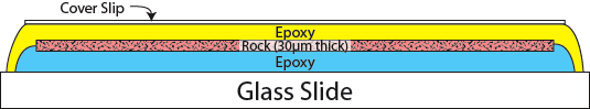
A thin section is a "slice" of rock brought to the thickness of about 0.03 mm. This thickness allows most of the
minerals present to be traversed by light and therefore, thanks to the use of the polarizing microscope, a whole series of investigations can be performed that allow their recognition.
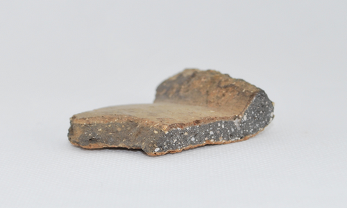
For the realization of a thin section we start from a fragment of stone which is representative of the petrographic characteristics of the rock to be examined and, if this is not possible, it will be necessary
make several thin sections for the study of the material.
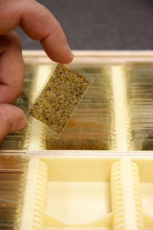
If the rock is inconsistent, it is necessary to start our entire preparation procedure with its impregnation and subsequent incorporation with epoxy resins.
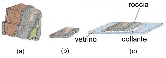
Once the resin has hardened, the cut can be made to obtain a brick of about 20x30 mm and the surface involved in the attachment to the glass slide is flattened.
The attachment to the specimen slide, suitably ground, also takes place by means of epoxy resin.
The next step is that of removing most of the sample, using special machinery, leaving only a small part of the rock that will be processed in the final preparation phase.
This phase involves the flattening of the sample, using silicon carbide powders of gradually finer grains, until the desired thickness is reached.
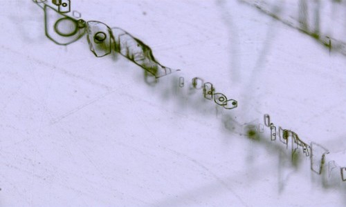
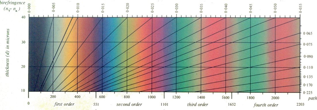
To be sure that we have achieved our goal, we analyze the thin section under a microscope by analyzing the colors that are not related to the color of the mineral (selective absorption of certain wavelengths), but to the interference of the two rays that have crossed the crystal with different speeds and they come out "out of phase". These colors are the INTERFERENCE COLORS, which depend on the properties of the mineral (in particular its "birefringence") but also on the orientation and thickness of the crystal crossed.
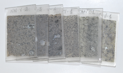
Once the desired thickness has been obtained, one then proceeds to affix a cover glass on the thin section, or proceed with the polishing phase for further analysis.
You can then proceed to the study of the thin section by means of the microscope, as per European standard UNI EN 12407.